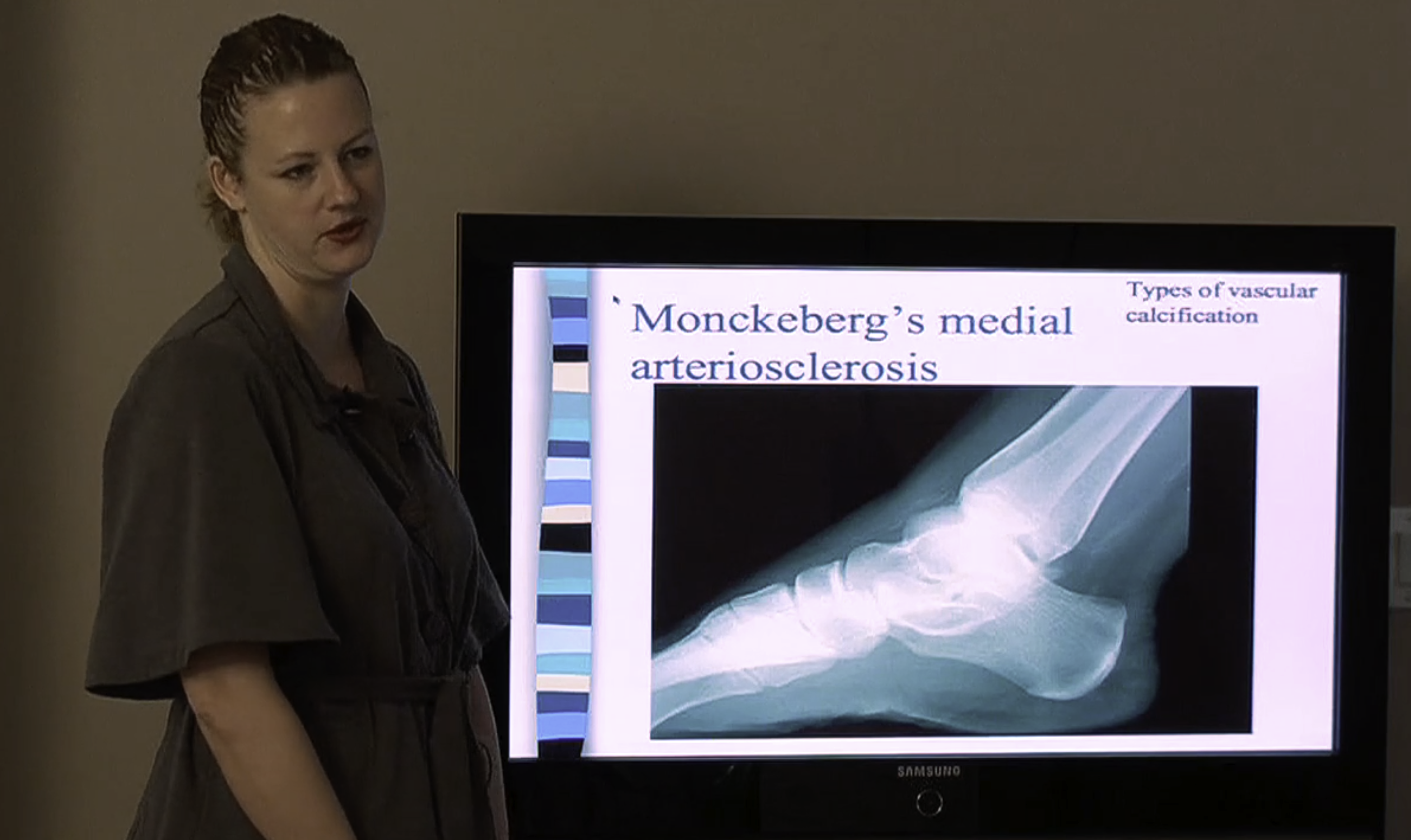
As presented by Dr. Osterhouse, Radiology in Practice teaches how to take and interpret radiographs, MRIs, CTs, and other diagnostic imaging tools, including the bone scan. In addition, the course provides a valuable review of spinal and extremity imaging for a doctor in general practice.
This hour teaches the participant about the presence and development of various pathologies, how they can be visualized with specific diagnostic imaging tools and explains x-ray findings in the diabetic patient. This course will enhance the student’s ability to take and interpret radiographs of patients who have diabetes. Dr. Osterhouse provides a review of diabetes, offering a typical patient’s history and examination findings. She offers radiographic examples and suggestions about how to use x-ray in the management of the diabetic patient. This hour provides a comprehensive review of the diagnostic imaging finding related to diabetes that will further the doctor’s understanding of this common condition.
Learning Objectives
The participant will be able to discuss what diabetes is and how it affects the nervous and skeletal systems. In addition, they will be able to explain how to utilize diagnostic imaging to appropriately diagnose and assist in the care of a diabetic patient.


